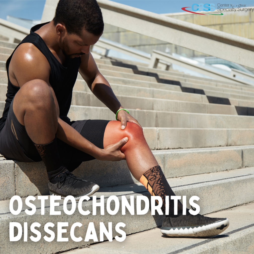
Osteochondritis dissecans (OCD) is a condition that develops in joints, most often in children and adolescents. This condition occurs when a small segment of the bone partially or fully separates from the end of the bone that forms a joint. This usually happens due to the lack of blood supply to the area. As the piece of the bone dies, and the cartilage covering it begin to crack and loosen.
In many cases of OCD in children, the affected bone and cartilage heal on their own, especially if a child is still growing. However, in adolescents, OCD can have more severe effects. The OCD lesions have a greater chance of separating from the surrounding bone and cartilage and can even detach and float around inside the joint. The most common location of OCD is in the knee at the end of the femur (thighbone). It can also affect other joints, such as elbows and ankles.

CAUSES
It is not known exactly what causes the disruption to the blood supply and the resulting OCD. However, osteochondritis dissecans has been linked to:
⦁ Repetitive trauma or stress on a joint, such as from playing sports
⦁ Genetic predisposition in some patients
SYMPTOMS
Pain and swelling of a joint — often brought on by sports or physical activity — are the most common initial symptoms of OCD.
Other signs and symptoms of this condition include:
⦁ Locking and “catching” of the affected joint
⦁ A “giving way” sensation in the affected area
⦁ Changes in the range of motion in the joint
DIAGNOSIS
No laboratory studies are indicated in the workup of OCD. Diagnosis is clinically made from the history with support from imaging studies which typically begins with plain radiographs and often progresses to magnetic resonance imaging (MRI) scans, to confirm the diagnosis and guide treatment. Computed tomography (CT) scanning may be helpful in preoperative planning and in guiding treatment when MRI is not available or is contraindicated.
TREATMENT
The treatment of OCD largely depends on the affected joint, the size, and the degree of separation. The management could be non-surgical or surgical. The healing of the OCD should be monitored with routine imaging
NONSURGICAL OPTIONS INCLUDE:
⦁ Rest
⦁ Modification of activities to reduce stress
⦁ Brace, boot, or cast to immobilize the affected joint
SURGICAL TREATMENT
Your doctor may recommend surgery if:
⦁ Nonsurgical treatment fails to relieve pain and swelling
⦁ The lesion is separated or detached from the surrounding bone and cartilage, moving around within the joint
⦁ The lesion is very large (greater than 1 centimeter in diameter), especially in older teens
The goal of the surgery could be to:
⦁ Stimulate an unhealthy area of the bone to heal
⦁ Secure a loose bone fragment to allow it to heal
⦁ Remove the osteochondritis dissecans lesion and reconstruct the cartilage (typically the last resort)
Surgical options include the following:
⦁ Arthroscopic subchondral drilling, to create channels that link the subchondral bone to the fragment
⦁ Arthroscopic debridement and fragment stabilization
⦁ Arthroscopic excision, curettage, and drilling
⦁ Open removal of loose bodies, reconstruction of the crater base, mosaicplasty for fragment healing, and potential replacement with fixation
⦁ Autologous chondrocyte transplantation
⦁ Radical removal of sclerotic bone with bone grafting of the defect and autologous chondrocyte transplantation (ie, sandwich technique)


comment (0)